Dr. Du Linna, a member of the research team led by Professor Cao Xuan at the School of Medicine/Institute for Advanced Study of Taizhou University, was invited to contribute a review to Journal of Hematology & Oncology (IF: 47.8), a leading international journal in hematology and oncology. The review, titled “Cellular Senescence and Regulated Cell Death in Cancer: Mechanisms, Cross-regulatory Networks and Therapeutic Implications,” has been published in the journal.
Dr. Du Linna of the School of Medicine/Institute for Advanced Study at TU and Zhang Yueyue, a graduate student at TU, are co-first authors of the paper, with Du listed first. Liang Ruohan, an undergraduate student at the School of Medicine, Associate Professor Zhou Jun, and Professor Xia Xianwu of Taizhou Municipal Hospital are co-authors.
Professor Cao Xuan of the School of Medicine/Institute for Advanced Study is the corresponding author, and Professor Wu Kongming of Tongji Hospital of Huazhong University of Science and Technology and Shanxi Bethune Hospital, together with Professor Liu Chibo of Taizhou Municipal Hospital, are the co-corresponding authors.
TU is listed first among the institutional affiliations in the paper; the first affiliation entry covers the School of Medicine and Taizhou Municipal Hospital, the university’s affiliated municipal hospital. This work was supported by the National Natural Science Foundation of China (Grant No. 82473945).
Faced with stress, the fate of a cancer cell is not simply a binary choice between survival and death: it may enter cellular senescence, a state in which it stops proliferating but remains alive, or it may activate one of several regulated cell death programs.
Cellular senescence can serve as an anticancer defense, as damaged or premalignant cells halt division and immune signals may promote their clearance. But senescence is a double-edged sword. Senescent cells remain biologically active and can release inflammatory factors, growth factors, and tissue-remodeling enzymes, giving rise to the senescence-associated secretory phenotype (SASP).
If these cells persist, SASP signaling can shape a tumor-favorable microenvironment that promotes immune escape, treatment resistance, or recurrence; some cancer cells may also escape the senescence-induced arrest and resume growth.
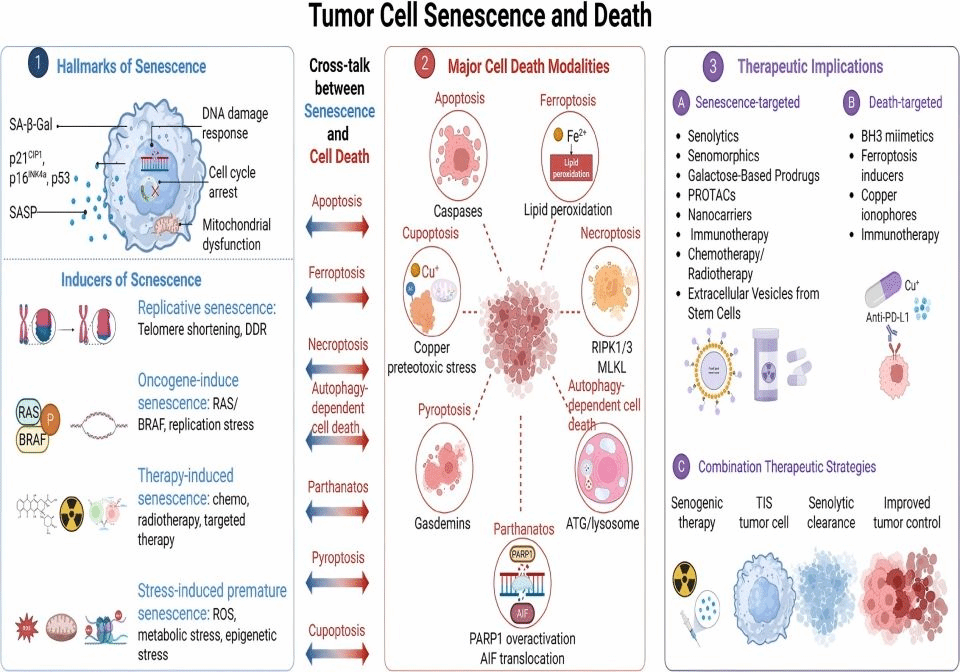
Schematic diagram of the interaction network between tumor cell senescence and death
The review distinguishes among senescence in premalignant cells, therapy-induced senescence in established tumor cells, and senescence of non-malignant cells within the tumor microenvironment, noting that these different contexts may lead to different outcomes.
It places cellular senescence and cell death within a single framework, explaining how the two jointly shape tumorigenesis and progression, treatment response, and recurrence. It maps the connections between them and the transitions that may occur between them in specific contexts, and examines the implications of these findings for clinical treatment strategies as well as the challenges involved in translating them into practice.
The review surveys seven forms of regulated cell death associated with cancer: apoptosis, ferroptosis, autophagy-dependent cell death, necroptosis, pyroptosis, parthanatos (PARP1 hyperactivation-mediated cell death), and cuproptosis. Their mechanisms differ, and so may their effects on tumor control and immune responses.
To explain how cell fate is determined, the review integrates evidence around a set of interacting regulatory hubs, including the DNA damage response (DDR), p53, mitochondria, autophagy, and epigenetic regulators. These systems jointly sense the type, intensity, and duration of stress and integrate information on the cell’s repair capacity, metabolic state, and microenvironment.
As a result, similar stresses can lead to divergent outcomes, ranging from adaptation or senescence to regulated cell death. Under certain conditions, the link between the two goes further: senescent cells can be driven into regulated cell death.
This framework suggests that therapeutic thinking should not stop at inducing senescence. One two-stage strategy now under investigation is to first drive tumor cells into senescence through anticancer therapy, and then selectively eliminate the persisting senescent cells using senolytic agents, suppress harmful SASP signaling using senomodulators, strengthen immune surveillance, or activate complementary cell death pathways.
Preclinical studies have already produced a number of examples of such combination regimens, for instance inducing senescence first and then applying pro-apoptotic therapy. However, most senescence- and cell death-targeted strategies in oncology remain at the preclinical stage, and the clinical experience available to date is not yet sufficient to establish senolytic therapy as a routine treatment for cancer.
Several key questions must still be answered before translation can move forward: which biomarkers can identify the patients and senescent cell populations most likely to benefit; when senescent cells should be cleared; how to avoid damaging both the senescent cells that serve beneficial physiological functions and normal tissues; and whether the relevant pathway connections are present in intact human tumors.
The authors call for biomarker-guided studies, tracking of the temporal changes induced by therapy, rigorous testing of the causal relationships underlying cell fate transitions, and spatial validation in tumor tissue. The central message of the review is that cellular senescence and regulated cell death should be studied as an interconnected, context-dependent network.
Only by clarifying the mechanisms, the timing, and the safety profile can combined therapies be optimized on this basis.
Article link: https://doi.org/10.1186/s13045-026-01847-x